VisionPACS® Advance
The power to transform images into decisions
After 17 years of research and development, after a few hundred million lines of code and after a few dozen versions, the VisionPACS® Advance workstation has reached the level of maturity necessary to be a leader in several countries. Today VisionPACS® Advance is taking on the challenge of becoming the world’s leading medical image processing software.
-
Description
VisionPACS® Advance is a scalable solution that starts with a basic version and allows you to add the options of your choice at a lower cost. Its intuitive, comprehensive and extremely powerful user interface (2D, 3D, Volume Rendering, MIP, MPR, Autobone, Vessel Analysis, etc.) makes this post-processing platform an essential tool for establishing a diagnosis. Its power and speed will allow you to save time and optimize the workflow in your radiology department or practice.
Database:
Local multi-modality database, multi-service DICOM Store, Q/R, WorkList, MPPS, DICOM J2000.
Display/edit DICOM tags.
Import and burn CD/DVD with integrated viewer.
Export images in bmp, jpeg, avi format…
Ergonomics and interfaces:
Quick and easy to get started.
Toolbar based on exam type and study type.
Image thumbnails for easy navigation.
Security:
Authentication by login and password.
Access control by list of rights and permissions.
Deletion and modification protected by password.
Controlled anonymization of exams.
Displaying and processing images:
Display and processing of images on one and two screens.
Support of a 3rd screen for the list and the report.
Measurements (angles, distances, surfaces, densities)
Annotations, Rotation of images, Graduated scale, Zoom, magnifier.
Cine mode.
Special processings:
Stitching and pasting of images.
Subtraction and addition of images.
Measurement: Cobb angle, TAGT and Coxometry/Goniometry.
Printing:
DICOM PRINT.
Automatic and manual film composition.
Real size printing.
Film composition Backup.
Customizing film composition with image areas of different sizes.
Printing on paper printer.
3D and post processing:
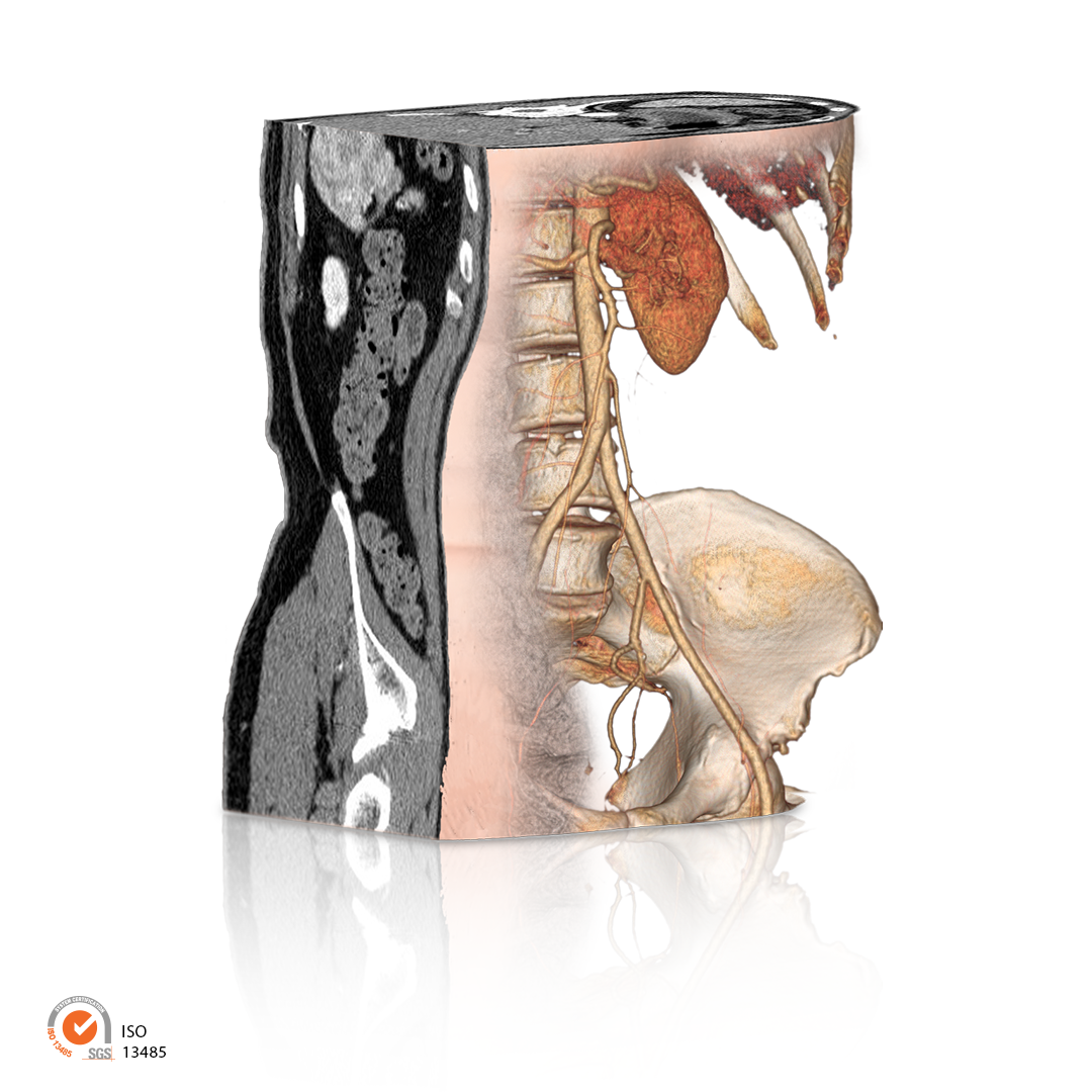
Real-time variable-thickness MPR reconstruction.
Reconstruction of derived series of variable thickness.
Orthogonal and curved MPR reconstruction.
2D oblique and double oblique reconstruction of variable thickness (MIP,
MinIP and AveIP)
3D reconstruction in VR/MIP/MinIP/AveIP.
Real-time premium 3D hardware acceleration.
Spherical shutter on 3D volume.
Manual segmentation of vessels and bone structures.
Oncological follow-up:
Oncological follow-up according to the RECIST 1.1 evaluation criteria.
Creation of special oncology reports.
Dental Study:
Creation of reconstructed series of dental cuts.
Creation of panoramic images.
Real size printing.
Study of multiparametric MR examinations:
Automatic detection of multiphase series.
Manual adjustment of pre-enhancement and wash-in wash-out ranges.
Display of the time/signal intensity line graph of a 2D or 3D ROI.
Chromatic display of Signal Enhancement Ratio “SER” (Signal
Enhancement Ratio )
Chromatic display of the Maximum Slope of Increase “MSI”
(Maximum Slope of Increase)
Chromatic display of Positive Enhancement Integral “PEI” (Positive
Enhancement Integral)
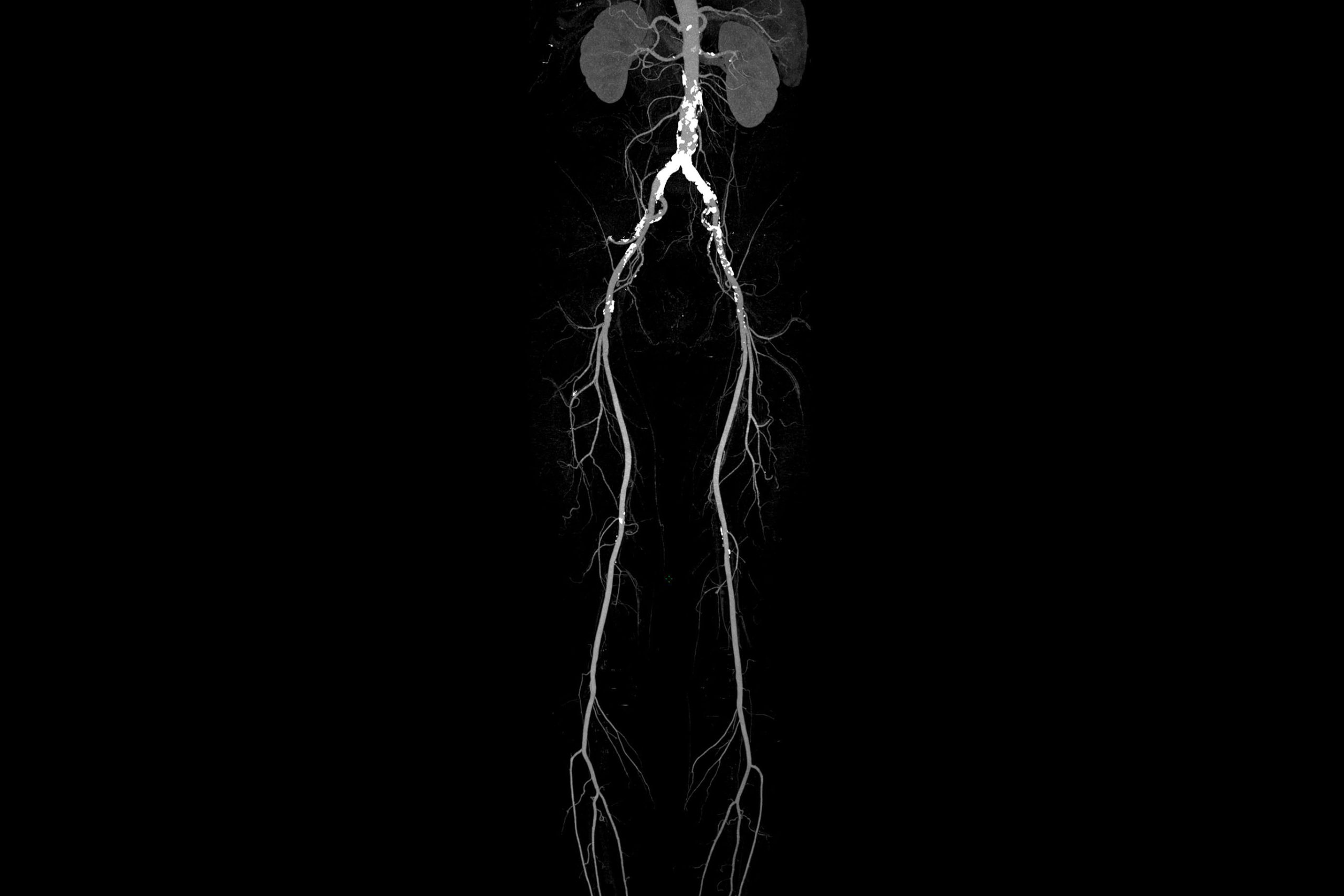
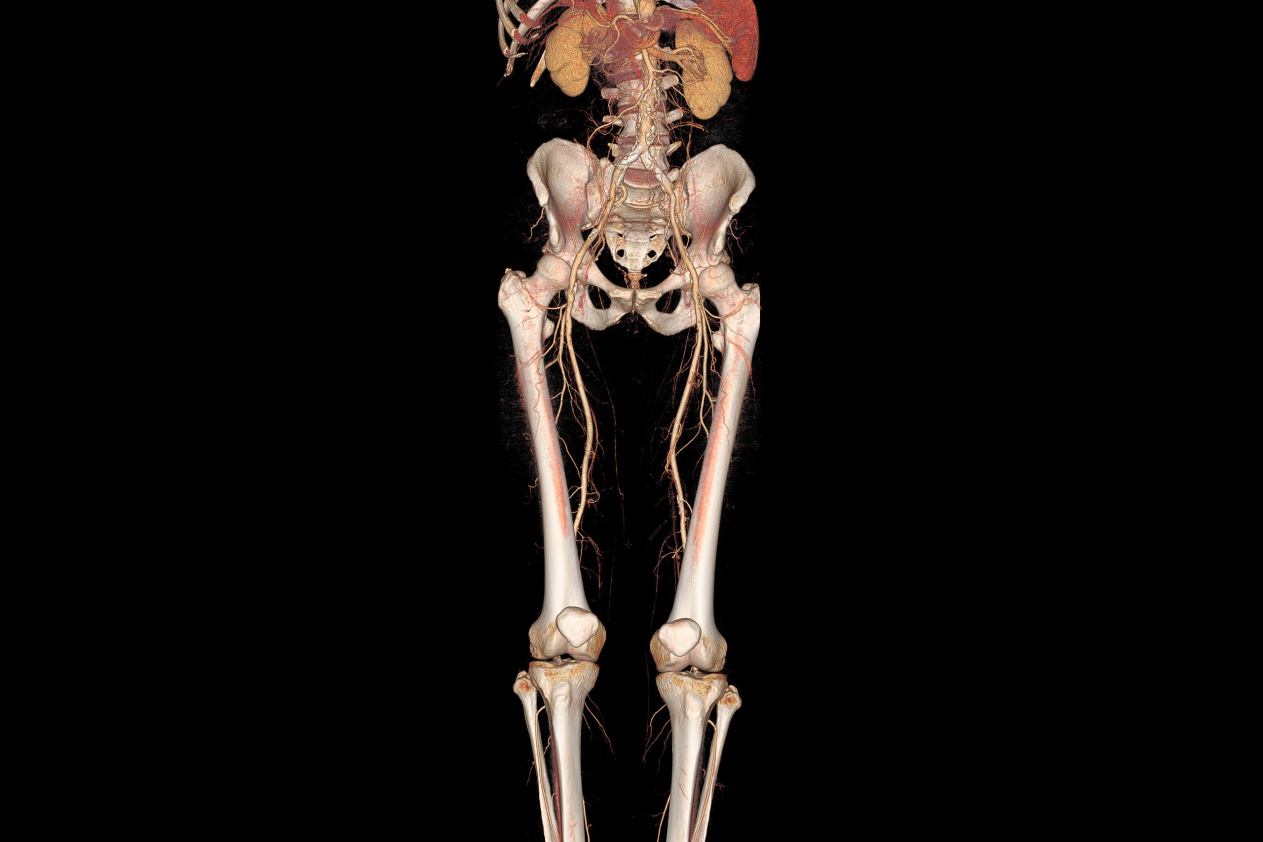
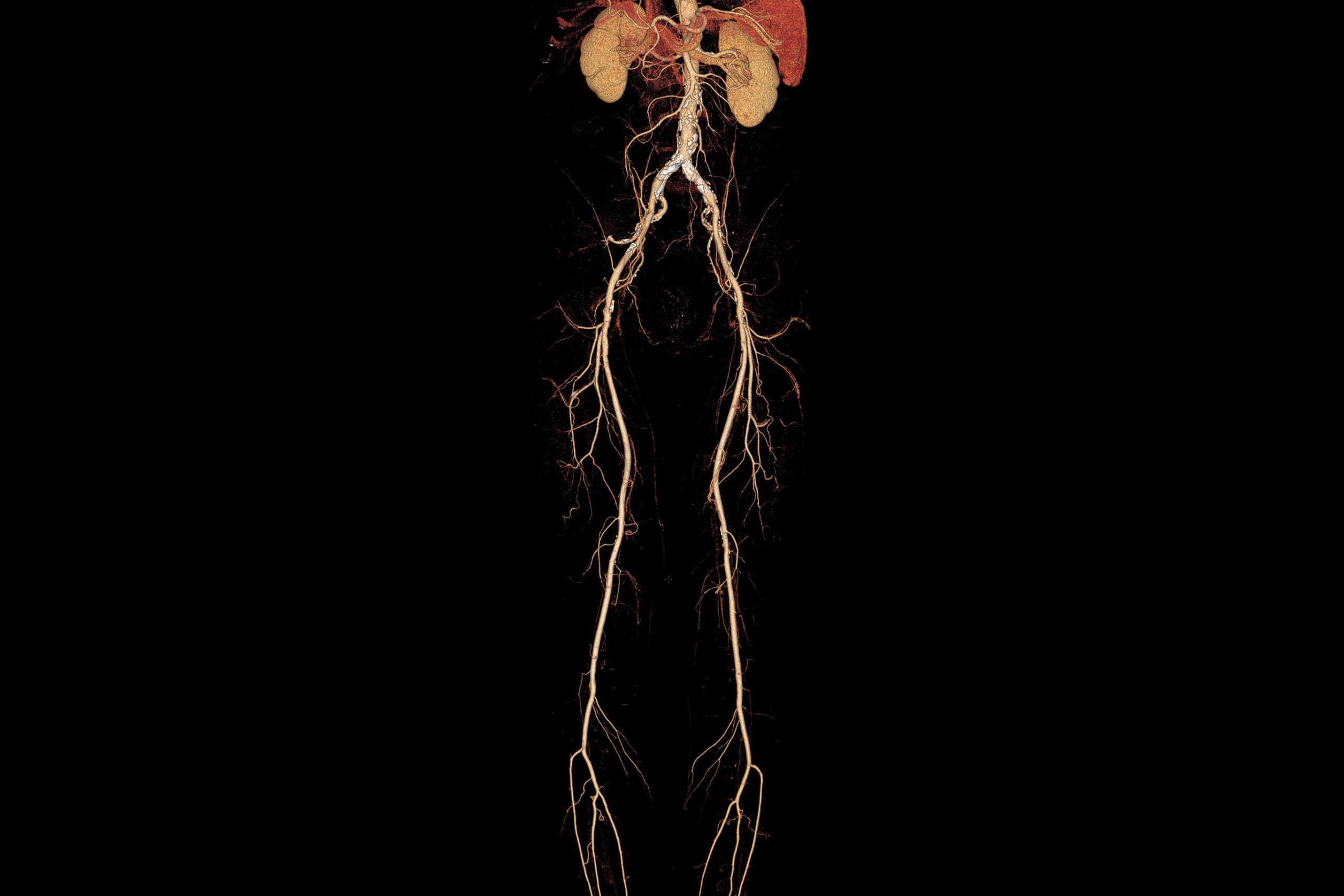
Advanced Vessel Analysis:
Bone segmentation and automatic vessel detection for examinations of the Thorax, abdomen and lower limbs (Autobone)
Detailed analysis of vascular structures.
Study of stenoses and aneurysms.
Tools for the automatic and manual improvement of vessel centerlines.
Creation of special vascular reports.
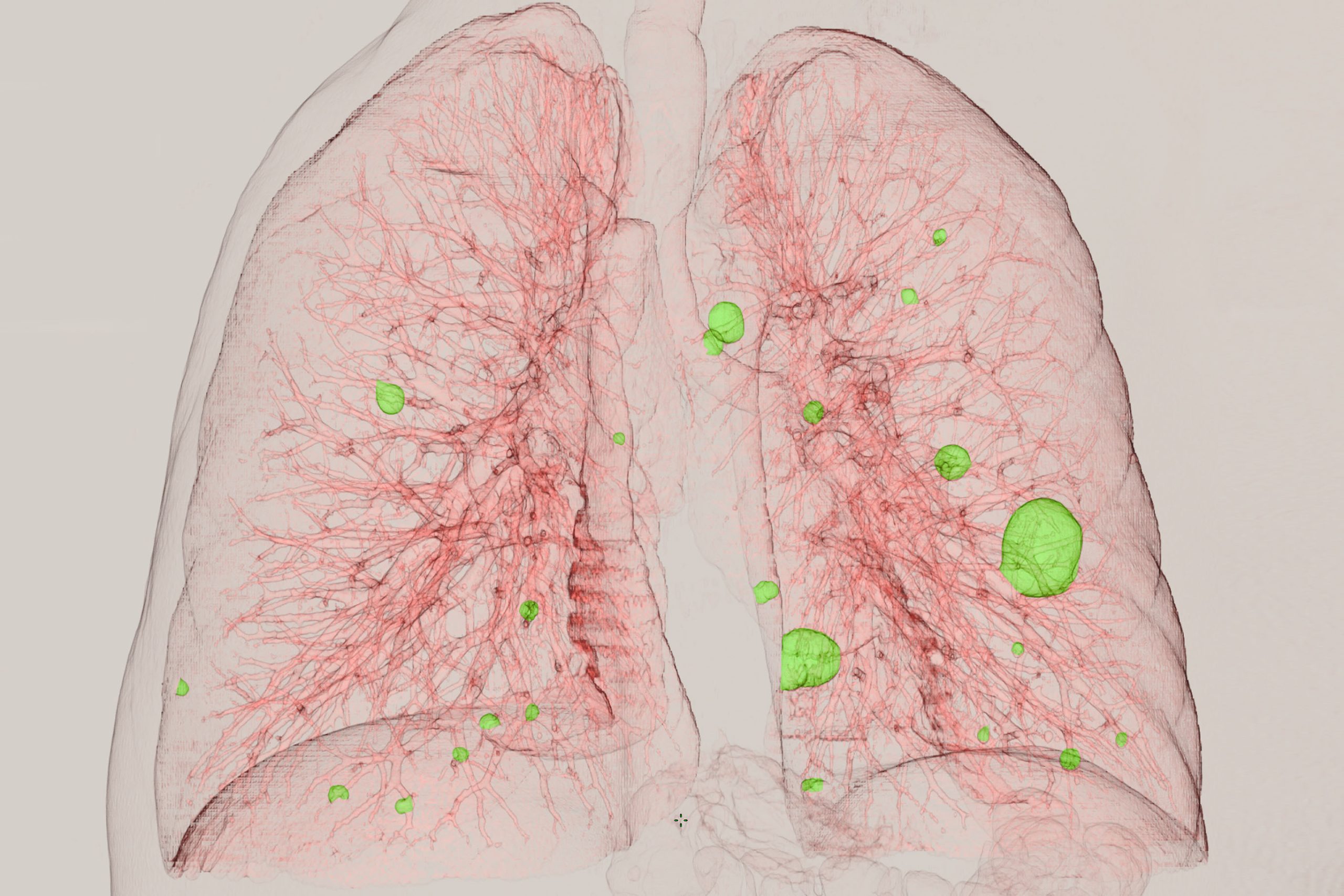
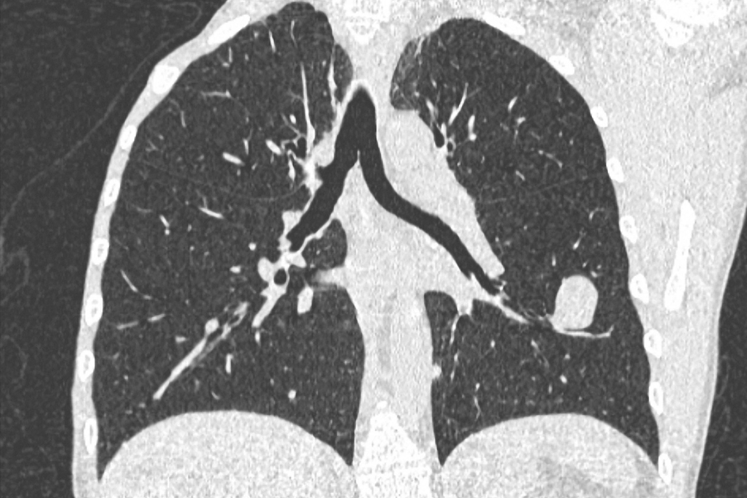
Lung study:
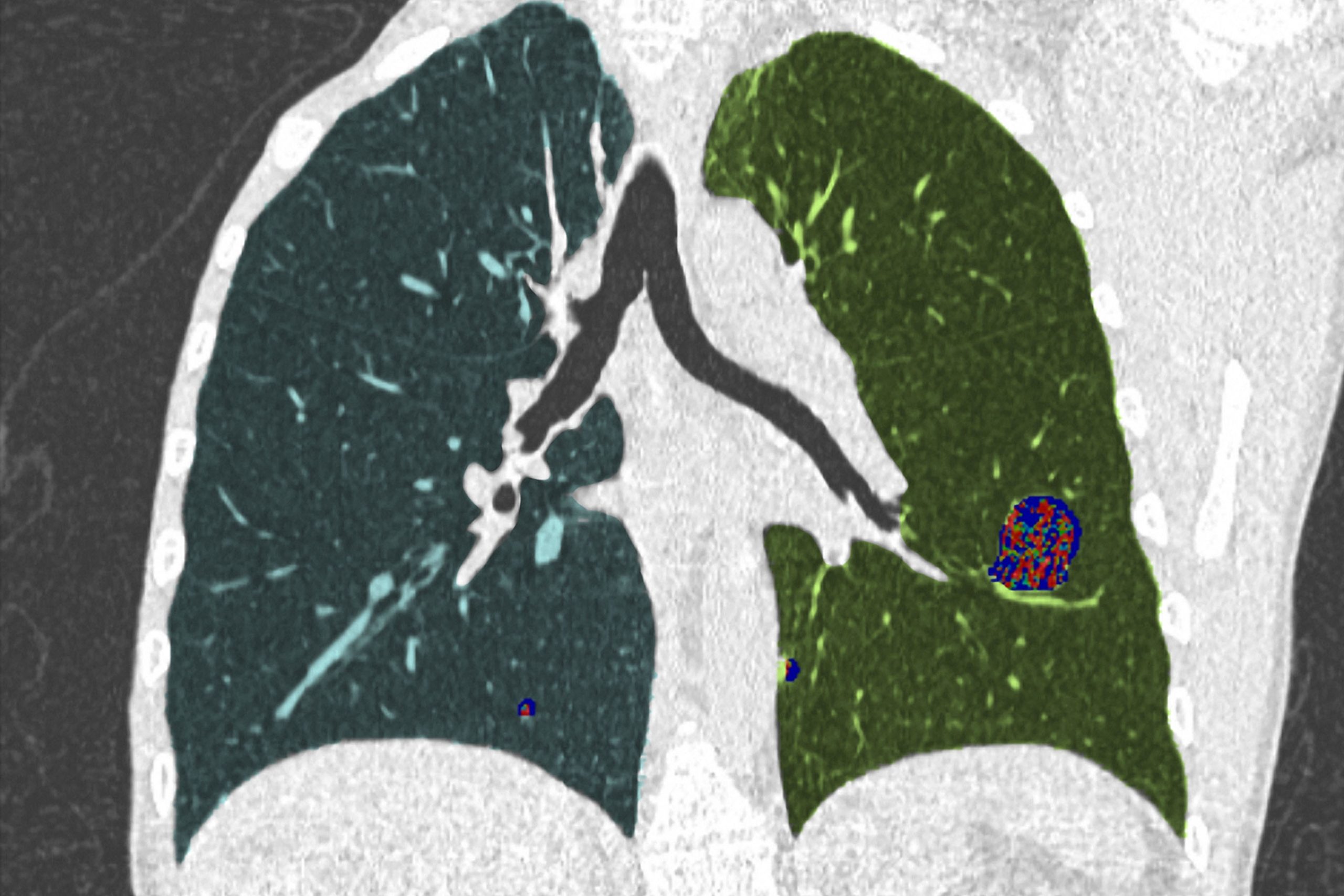
Automatic segmentation and detection of right and left lungs with volume calculation.
Calculation of pathological volumes.
Automatic detection of lung nodules.
Creation of special lung reports.
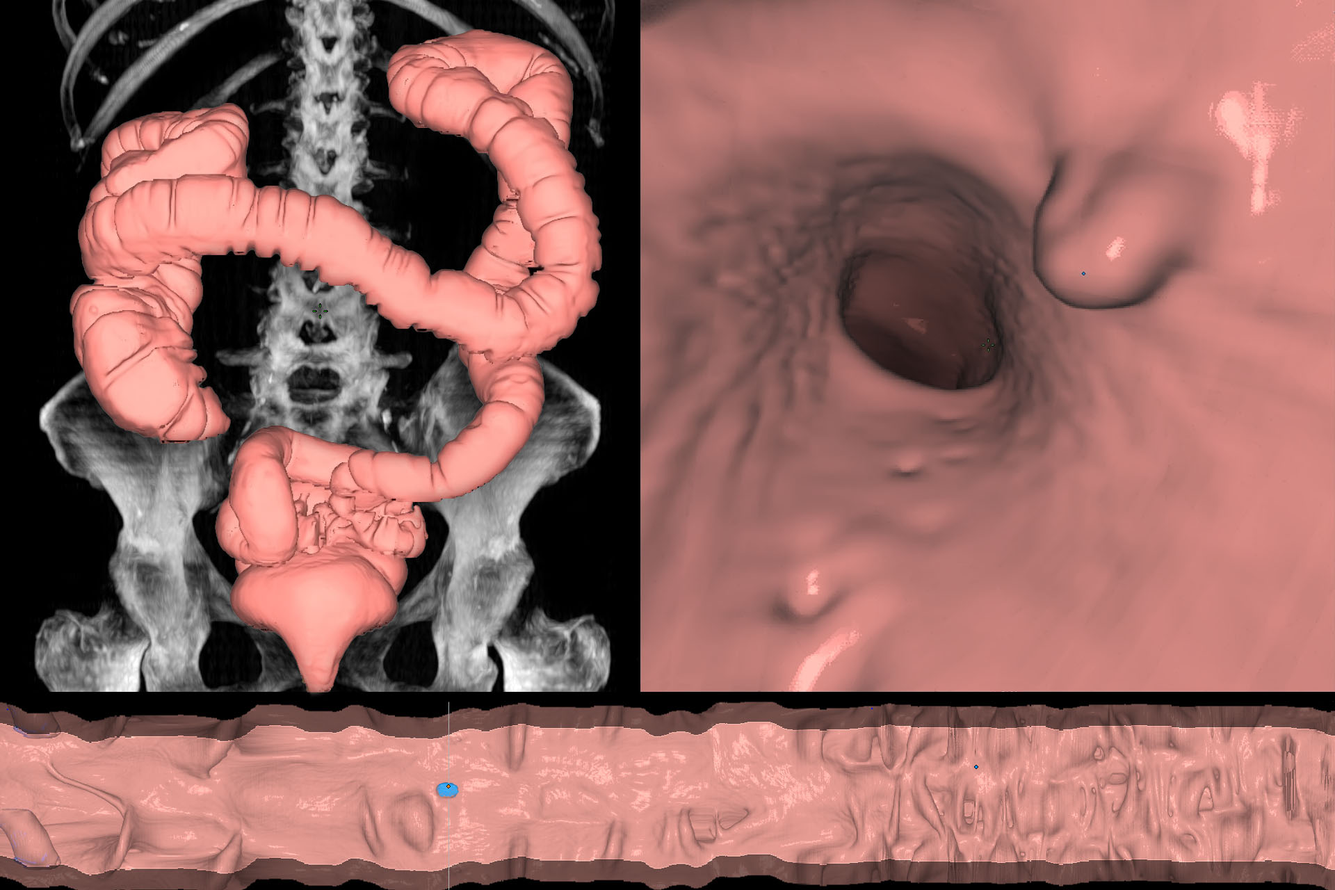
Colon study:
Automatic colon detection in supination and pronation.
Synchronized multi-view display of series (Supine/Prone)
Synchronized endoscopic navigation with 360° vision.
Automatic detection of polyps and suspicious tissue.
Spread out view of the colon.
Creation of special colon reports.
